Importance of Radiographs for Foot Balance
There are many opinions on how to “correctly” balance the foot with the naked eye. Many farriers are trained to look for certain landmarks of the hoof to guide how to trim/shoe that are learned from generations of very observant people. Some of those landmarks include the widest point of the hoof or frog, coronary band angle, duckets-dot. Advancements in technology and shared learning in the podiatry world have given us many articles referring to these anatomical landmarks and different ways to apply a trim or shoe. Fortunately, most of these methods do overlap. After all, every hoof care professional’s goal is to keep the horse sound and comfortable. Although methods may vary from one farrier to another, alternate factors that the farrier cannot always control should be taken into consideration. Genetic predispositions, wet environment, and medical abnormalities may skew traditional landmarks that used to be so reliable. This is where a great farrier-owner-veterinarian relationship can thrive to help your horse. Radiographs cannot lie.
Proper foot radiographs can guide your veterinary/farrier team to help your horse. There are many measurable guidelines that we can use to help decide on trim, shoe selection/placement, and even possibly a medical diagnosis to explain why the horse is not sound. Radiographs early on can help guide your farrier for proper balance to possibly prevent soundness issues down the road! That’s the power of a great veterinary farrier team. Below I have included two images of the same horse to explain just one of the many measurable guidelines we use with digital radiography: palmar angle.
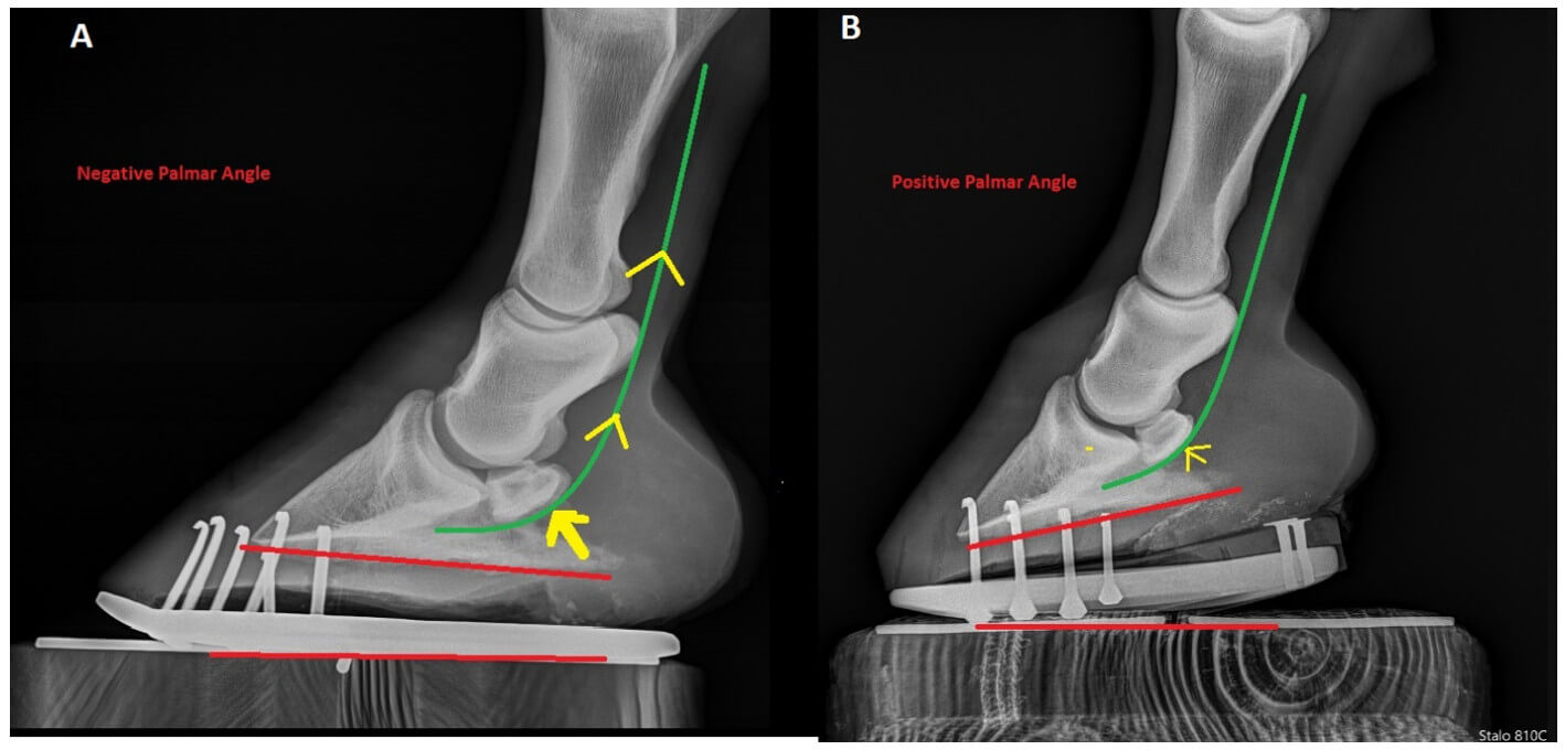
The red lines in the figure show the relationship between the bottom of the coffin bone and the horizontal ground surface. Figure A shows a negative angle in relation to the ground, whereas Figure B shows a positive angle. The green curved line represents the Deep Digital Flexor Tendon (DDFT) that runs below the navicular bone and attaches to the coffin bone to be able to flex the leg. The negative angle applies more tension on the DDFT, thus more force on the navicular bone. This angle over time can cause pain on the navicular bone and surrounding structures. Figure B represents the same foot trimmed with a shoeing package applied. Now the angle is positive, thus relieving pressure on the navicular bone and easing the force required for the foot to breakover forward. Unfortunately, many times if you manipulate one aspect of the foot, another may be compromised. In this case, heel elevation can cause the heels to be overloaded and crushed. This is why you may see one farrier do one package while the other may choose something different. There are numerous ways to achieve the same goal, and each way is just trying to minimize the side-effects of another. Just because each farrier may do something different, doesn’t always mean it’s wrong, as long as there was a thought process behind it.
In conclusion, radiographs are always a great start to help your veterinarian and farrier achieve their goals with your horse. Tennessee Equine has a great group of veterinarians motivated to work with your farrier. I personally have strived to make relationships with as many farriers as possible in the area and to continue my education in the podiatry field. I look forward to the opportunity to meet your horse with your farrier to get our hands dirty to develop the best option for your horse’s feet. No Foot, No Horse!