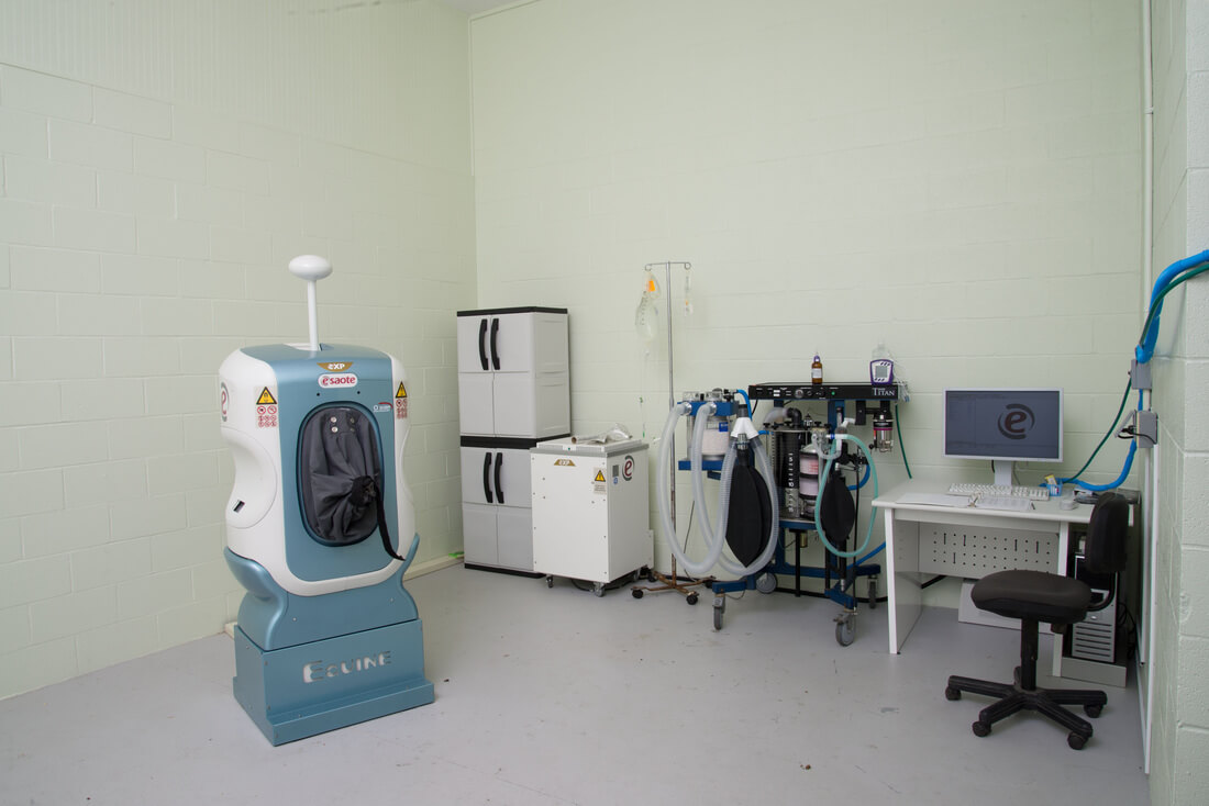
Esaote O-scan 0.31 Tesla MRI
Advances in equine imaging modalities have increasingly allowed us to diagnose lameness problems more completely and accurately than ever before. There are still some problems, however, that cannot be fully evaluated by radiographs, ultrasound, or even bone scans. Here at Tennessee Equine Hospital, we offer the latest in MRI systems to provide the most advanced diagnostic imaging for your horse.
Magnetic resonance imaging utilizes magnetic forces to align the protons in the body, which then transmit a signal to the computer that creates an image of the structure. Traditional high-field MRI systems require a special environment to protect the machine and people from the magnetic forces. Our system, the Esaote O-scan 0.31 Tesla, is a low-field scanner, which allows us to have the machine right in the clinic! This MRI system provides the high-field quality images, and is the fastest low-field scanner available. MRI images are obtained by looking at “slices” of the structure. Our system is able to evaluate down to 0.6 mm thick slices. This means anatomical structures are assessed in incredible detail in a 3-D manner. We are able to evaluate the lower limbs from the hocks and knees down to the hoof. All of our MRI images are sent directly to a radiologist who specializes in reading veterinary MRI scans. A thorough report is available in 24-48 hours in most cases, so treatment options can be determined quickly.
|
|